Home/Wellness Zone/Sakra Blogs
24th Feb, 2017
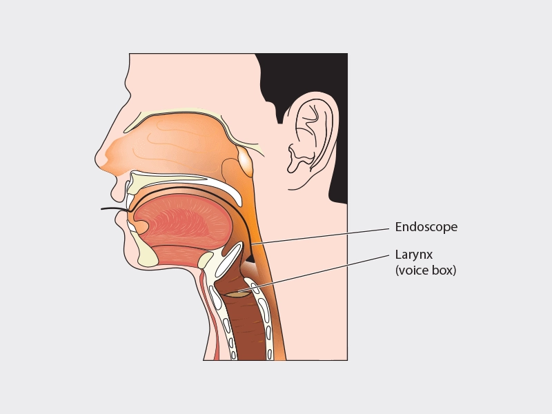
Head and neck cancers arise in multiple sites and sub sites, mostly detected so late that the oncological treatment encompasses extensive surgery and post-operative functional defects to the individual causing significant cosmetic changes, difficulties in swallowing, chewing, breathing and speaking. Hence early detection of these tumours in one of the main factors in treatment success.
Efforts to achieve the earliest detection of malignant disease have led to the development of new endoscopic examination methods. Endoscopic examination has hence become a corner stone for the diagnosis and treatment of cancers of the upper aero digestive tract. However, a laryngoscope with conventional white light (CWL) has technical limitations in detecting small or superficial lesions on the mucosa. Narrow band imaging especially combined with magnifying endoscopy (ME) is useful for the detection of superficial squamous cell carcinoma (SCC) within the oropharynx, hypo pharynx, and oral cavity.
Narrow band imaging (NBI) is a novel endoscopic technique enhances the diagnostic sensitivity of endoscopes for characterizing tissues by using narrow-bandwidth filters in a sequential red, green and blue illumination system. It exploits the neoangiogenetic pattern of vasculature, hence enabling to differentiate between benign & malignant/ premalignant condition. This filter cuts all wavelengths in illumination except two narrow wavelengths. One of these wavelengths is of 415 nm which corresponds to the peak absorption spectrum of haemoglobin to emphasize the image of capillary vessels on surface mucosa. Superficial lesions are identified by changes in the colour tone and irregularity of surface mucosa during endoscopic examinations.
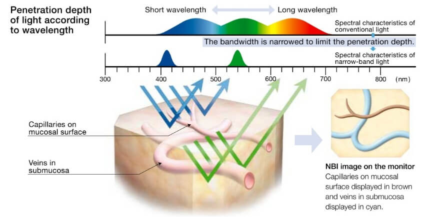
NBI is a non-invasive technique that can be carried out in the outpatient clinic without need of general anaesthesia. NBI system consists of the same components as conventional video endoscopic systems - light source, camera unit and camera head or chip equipped video endoscope. The blue filter enhance the image of capillary vessels (IPCL - Intraepithelial Papillary Capillary Loops) on mucosal surface. The 540 nm wavelength light penetrates deeper and highlights the sub mucosal vascular plexus. The reflection is captured by a charge coupled device chip (CCD), and an image processor creates a composite pseudo colour image, which is displayed on a monitor, enabling NBI to enhance mucosal contrast without the use of dyes.
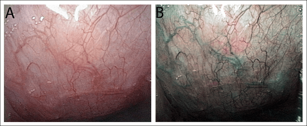
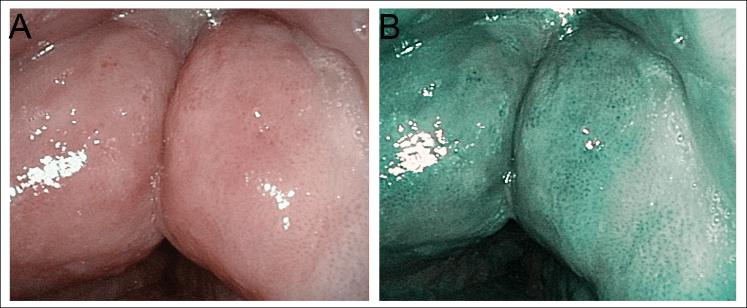
Healthy nasopharyngeal mucosa in white light (A) and NBI (B).
Submucosal vessels are visualised with greater contrast.
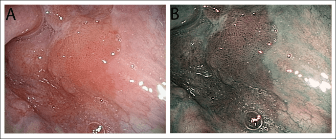
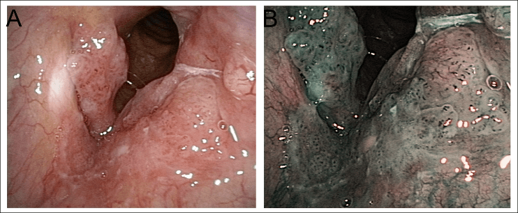
Dysplasia in Vallecula in white light (A) and NBI (B).
Submucosal changes for dysplasia, Carcinoma in situ can be appreciated as brown dots irregularly distributed in a well demarcated area. Hence NBI has proved useful in targeted biopsies of suspected areas and to determine limits of resection.
For follow up post Radiotherapy
NBI has high sensitivity (91.3 - 100%) and specificity (91.6 - 98%) in Head and Neck cancer follow up patients.
Supraglottic spread (of Glottic carcinoma) well appreciated on NBI.
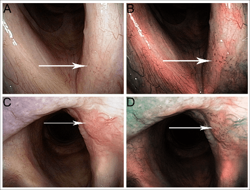
Dysplastic changes; brown dots on NBI
LIMITS OF NBI: As it is dependent on direct visualisation of the mucosa, conditions that prevent direct visualisation can alter the results obtained for example, pooled saliva, sticky mucus, or verrucous lesions where the epithelium has a layer of hyperkeratosis which prevents visualisation of the submucosa. Laryngeal papilloma is another entity where NBI cannot discriminate between benign & malignant lesions.
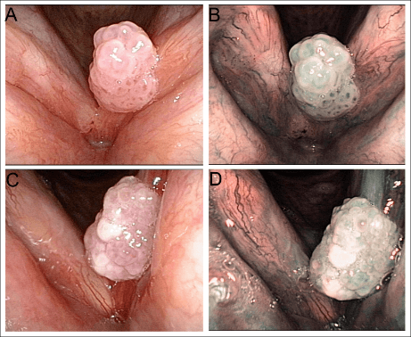
Laryngeal papilloma
NBI has proved to be a useful screening tool aero digestive malignancy, as well as in the pre-operative, intraoperative and post-operative staging of patients affected by squamous cell carcinoma in the head and neck area. Hence NBI is increasingly being used in otorhinolaryngology as a convenient screening method for detection of new diseases, but also for follow-up of patients after treatment for head and neck malignant tumours, when early detection of possible recurrence is crucial.
Enquire Now
